메트로아이센터 로고
- 스마일(SMILE)
- 스마일(SMILE)이란?
- 김명준원장 스마일 체험기
- 김종진원장 스마일 체험기
- 스마일 수술(실제) 비디오
- 올레이저 라식
- 올레이저 라식 vs 일반 라식
- 올레이저 라식 종류
- 라섹(넌터치)
- 라섹(넌터치)이란?
- 라섹(넌터치) 종류
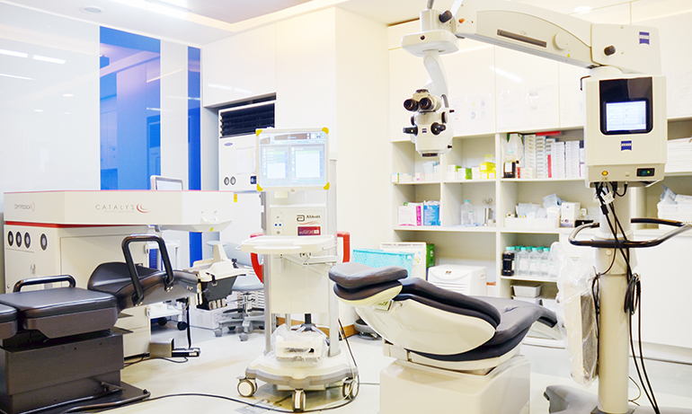
- 백내장 수술
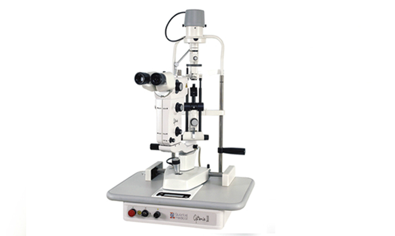
- 수술방법
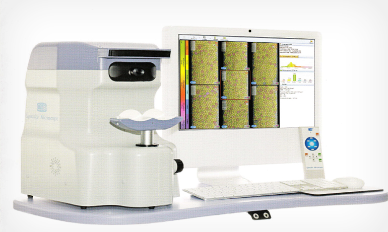
- 첨단장비의 ‘특별함’
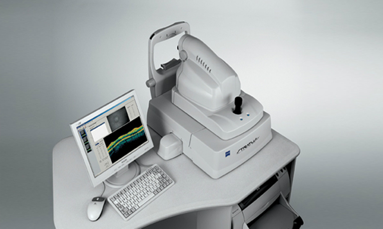
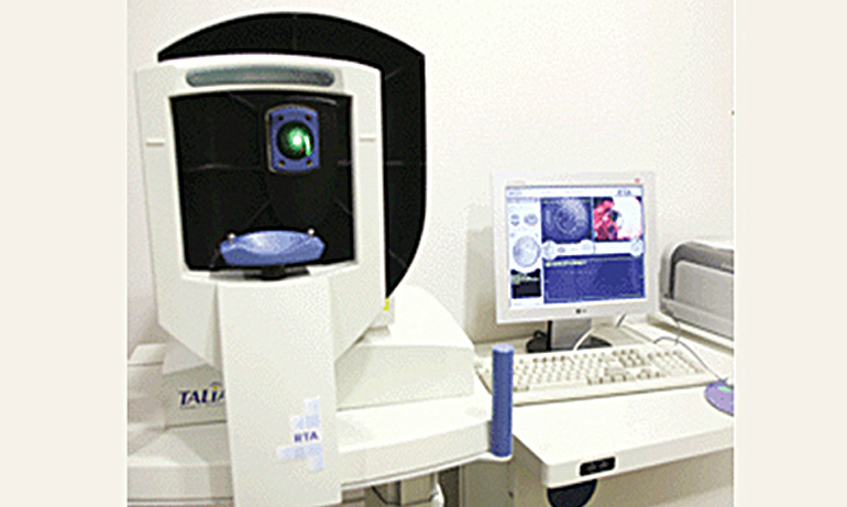
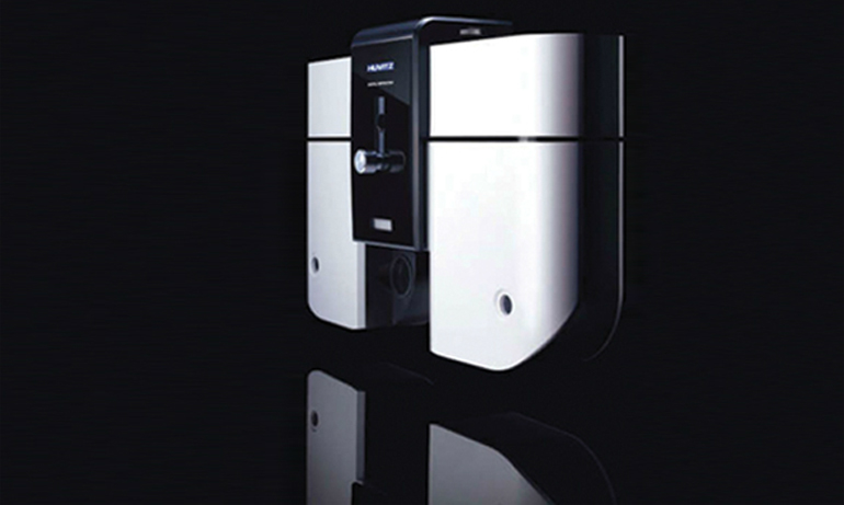
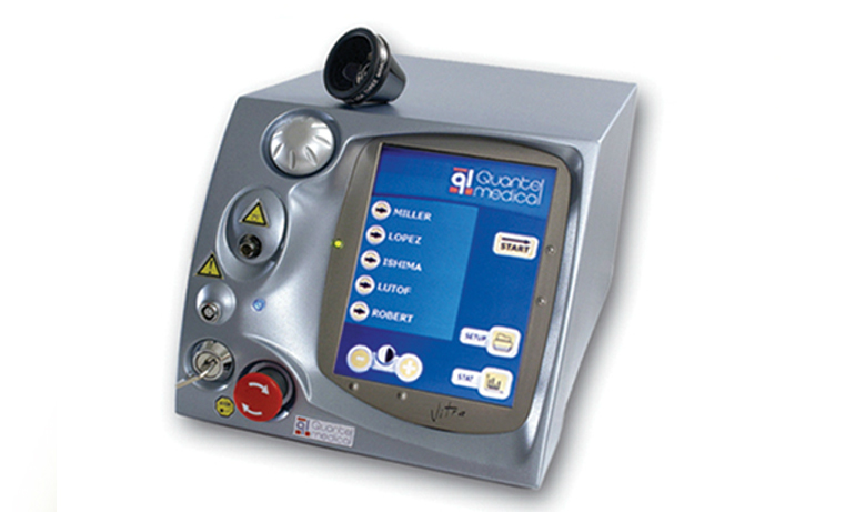
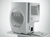
- 백내장 수술장비
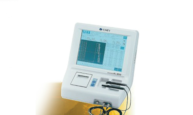
- 백내장 검사장비
- 수술 후 주의사항
- 백내장/노안 교정렌즈
- 교정렌즈 선택 길잡이
- 난시가 없거나 적은 경우
- 난시가 많은 경우
- 굴절 백내장 수술
- 노안과 백내장수술을 동시에
- 노안(40대이후)
- 노안이란?
- 노안 vs 근시
- 노안의 수술적 치료
- 노안의 비수술적 치료
- 40세이후 시력교정후기
About Us
Metro Eye Clinic is regarded as one of the leading eye care specialists in Deagu, South Korea. Our surgeons have helped thousands of people enjoy better vision. We are committed to excellence because the world turns to us for answers, and our patients turn to us for the best eye care treatment available.
Advanced
Technology- Metro Eye Clinic invests in the very latest laser, surgical and pre-op testing equipment available on the market. From LASIK & LASEK to cataract surgery and to the treatment of vitreo-retinal diseases, Metro Eye Clinic invests in the most high-tech equipment available. Our doctors are always up-to
up-to-date on the newest and best treatments and technologies.
Learn more about our newest lasers and other technologies by visiting our technology page.
Experienced
SurgeonsThe surgeons at Metro Eye Clinic are among the most experienced and well-trained in the entire
country. Together they have performed thousands of surgical and laser procedures.
They have taught courses to other doctors at major national and international meetings of ophthal-mologists. Our surgeons are fellowship-trained, having completed extensive training as eye surgery
specialists.Metro Eye Clinic is recognized as a center of excellence by major pharmaceutical and medical
device manufacturers. Our surgeons are continuously sought out by manufacturers to conduct
clinical researches and trials on new medications and technologies.
Dedicated
Surgery CentersMetro Eye Clinic has been awarded the certificate of LASIK/LASEK Clinic issued by the Korean
Ophthalmologists Association’s LASIK/LASEK committee. The award means that the surgery centers
has met nationally recognized standards for quality care. In order to achieve accreditation, the
surgery centers underwent an extensive on-site survey of its facilities and services. The survey team, composed of experienced ophthalmologists and administrators evaluated all aspects of patient care.Metro Eye Clinic also utilizes affiliated laser centers specially designed for vision correction.
The laser centers are constantly monitored to maintain strict humidity and temperature levels, which are imperative for the equipment to work properly.
Our Culture
- Every member of our Metro Eye Clinic team is dedicated to providing excellent patient care.
Our company motto is “Quality Patient Care First”, and we will work hard to make you feel like
you are our priority throughout every step of your care with us at Metro Eye Clinic.
What is the difference between LASIK and LASEK?
LASIK procedure doesn’t take up a lot of time.
Depending on the person, some people will get over the discomfort within 3 to 4 hours. Patients will have to use eye drops that they prescribe for about30 days. The aftermath varies from person to person but there is not a bigdifference.The day after the procedure about 90% of your vision has been cured.So, you can go back to your daily activities the following day. Also, patientshave less post check-ups than LASEK. It has little to no influence from sunlight.
LASIK is more expensive than LASEK due to the extra equipment that are used. LASIK procedure would be ideal for busy workers who would have go back to their daily life immediately, people who drive every day, or patients who willhave difficulty coming in for post check-ups.
LASEK and LASIK procedures are about thesame time.
But, patients of LASEK may feel more discomfort afterwards than the LASIK procedure. This discomfort feeling may last 2 to 3 days and patients will have to use eye drops for 3 to 6 months. The use of eye drops varies from person to person. Some patients may only need the eye drops for 3 months whereas others may need it for 6 months. The negative part to this is patients have to be out of sunlight for about 3 months. Patients must be extra careful with exposure to sunlight. This all varies from patient to patient. After the procedures, the amount of pressure that is put into your eyes will be relieved immensely.
Since there is a difference from patient to patient, it would be best to decide which procedure after going through multiple exams and consulting with a doctor.
This would lead to your next question,
why Metro Eye Clinic instead of other places?
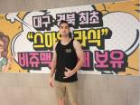
Aren’t they all the same? Actually, this is not true. Metro Eye Clinic has done the most vision correction procedures in all of Daegu and
Gyeongbuk region. To add onto their credibility, they have not any medical accidents that have occurred within the last ten years.
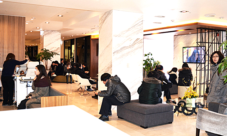
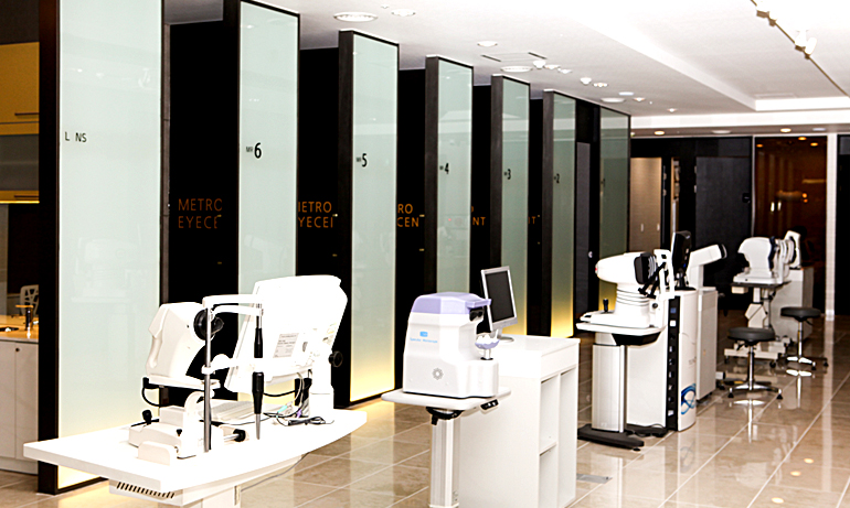
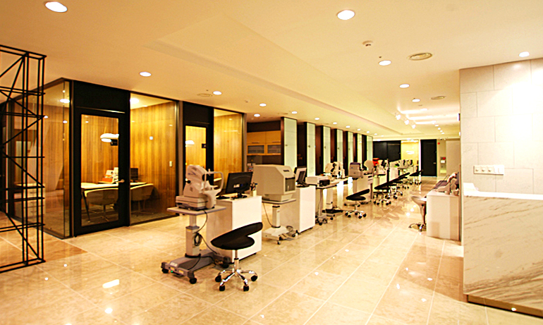
Metro Eye Clinic also uses the most state of the art equipment in comparison to other clinics. Different equipment is used to fully
check patients’ eyes. These equipment will examine the patients eye condition to give the best analysis before the LASIK or LASEK
procedure. Since everyone’s eyes are very different, these equipment can cater to virtually everything for the doctor to have a better
outlook and procedure that is customized for each patient. All of this equipment is from credible manufacturers internationally.
With this state of the art equipment, the price is actually a lot lower compared to other places around the region. The doctors have a lot of experience with these procedures, there are a lot of equipment to examine patients to the fullest extent, and over 100 doctors also received this procedure from the doctors at Metro Eye Clinic.
To give assurance to the patients, all of the doctors at Metro Eye Clinic received vision correction procedures from Metro Eye
Clinic. If it wasn’t safe, would these doctors have received this operation?
Since all of the doctors at Metro Eye Clinic have experience as a patient receiving the treatment, they are able to explain it better from
the patient’s point of view. Whatever uncertainty you may have felt, these doctors will be there to answer them and assure you that you
are under the right hands.
Surgery Procedure
Surgery can take place on the same day if you register in the morning.
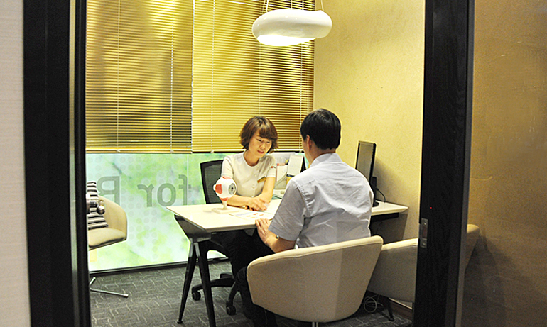
After registration, you will go through various check-ups to get a full understanding of your eye's condition.
The doctor will answer all of your questions regarding the surgery and also brief you on your condition.
the doctor will meet with you to advise which vision correction surgery would be proper for your condition.
Also, the doctor will take a closer look at your eyes after reviewing the check-ups that were done beforehand.After consulting with the doctor, the surgery will take place.
You must come back for follow ups for the doctor to see how your eyes are healing.
All of these check-ups are included in the surgery price.
How to get there
- Car: Novotel Daegu building. Parking is available using the same garage for Novotel and Daegu City Center.
When you park, take the B Elevator to the 6th floor. - Subway: Get off at Joongangro Station (Daegu Subway Line 1) and go to Exit 15.
You will be right in front of Daegu City Center/Novotel. Use the B Elevator to the 6th floor.
Pricing info on Vision Correction Surgery
- LASEK Price is about 1,000,000 won
- LASIK Price is about 1,500,000 won
- The price includes post check-ups (6 months) to see how you are healing.
Business hours
- Monday, Tuesday, Thursday, Friday, Saturday 09:00 ~ 17:00
- Wednesday 14:00 ~ 17:00
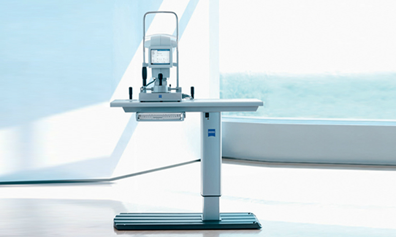
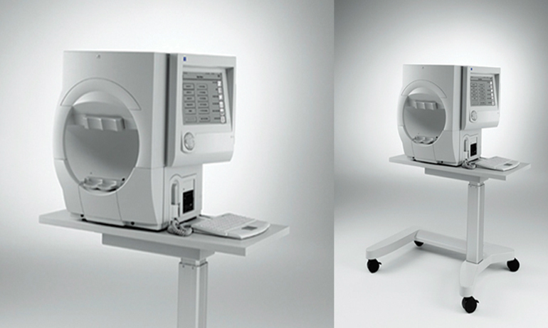
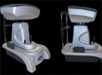
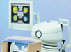
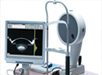
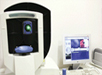
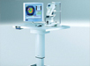
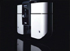
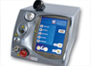
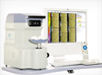
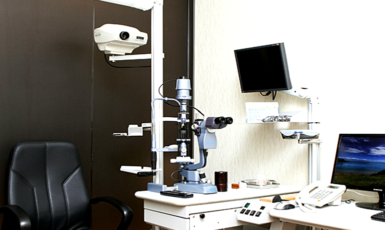
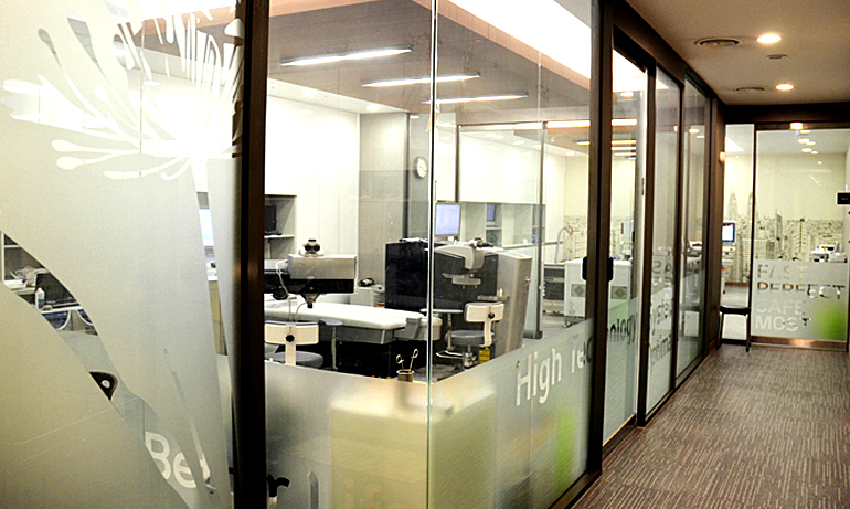
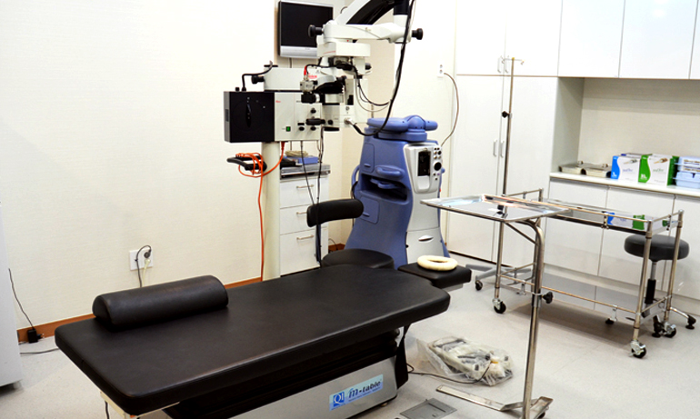
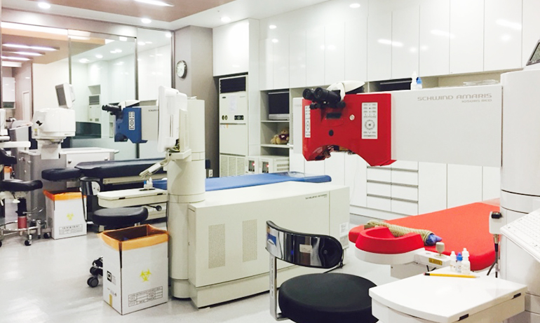
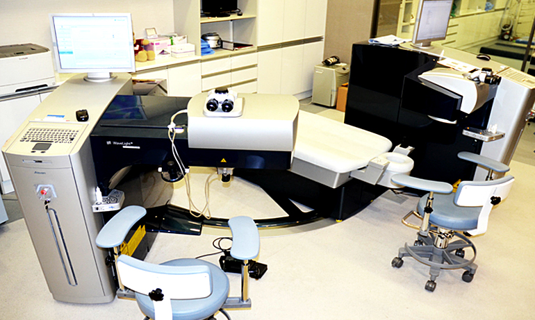
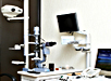
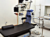
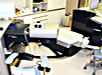
LASIK & LASEK laser surgical equipment
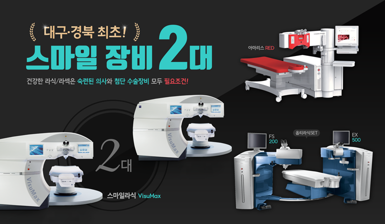
For more information on the laser surgical equipment
EX500 FS200 http://www.alconsurgical.com/
AMARIS 1050RS http://www.eye-tech-solutions.com/en/home/treat/schwind-amaris-models/schwind-amarisr-1050rs/
ZEISS <SMILE> https://www.zeiss.com/meditec/int/products/ophthalmology-optometry/cornea-refractive.html
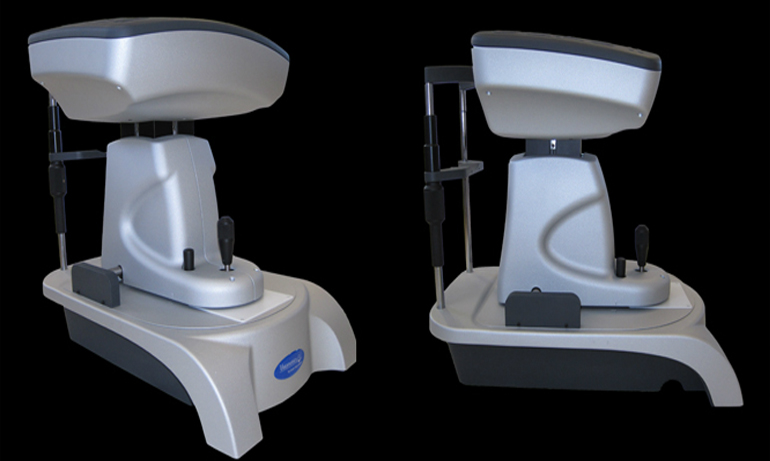
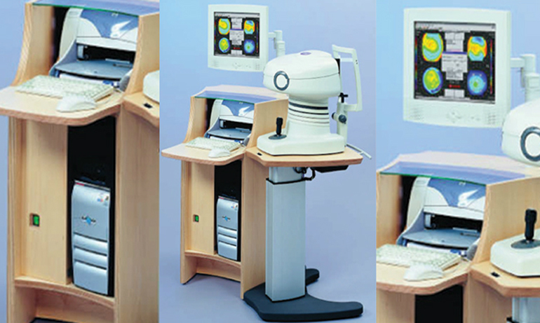
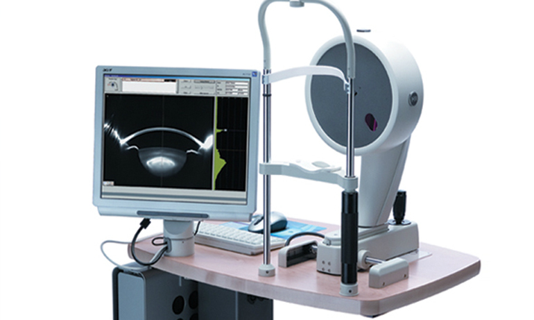
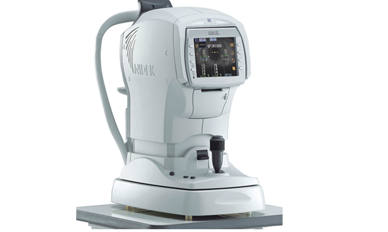
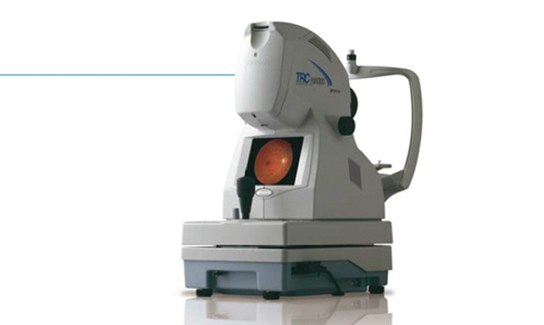
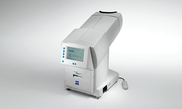
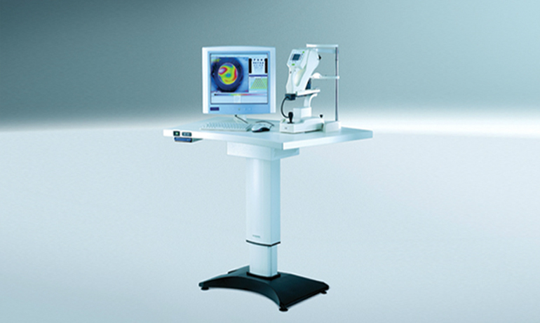
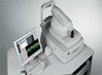
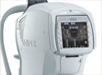
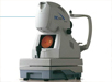
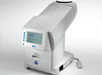
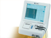
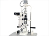
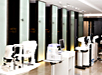
Pre-op testing equipment
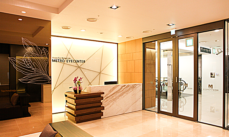
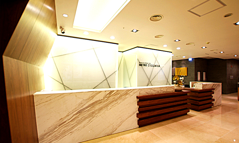
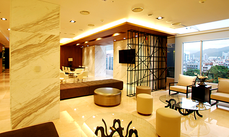
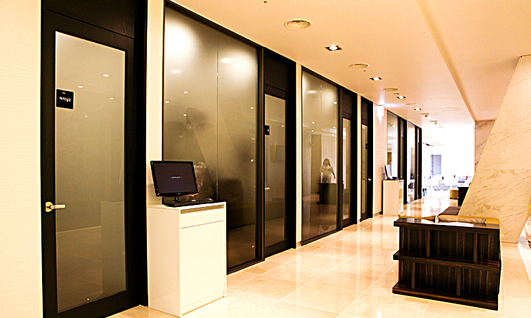
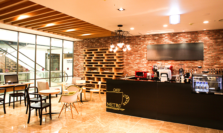
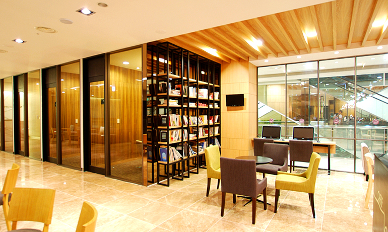
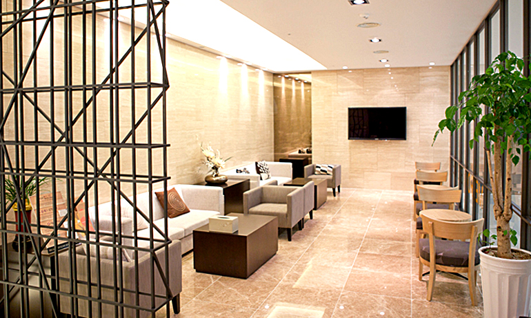
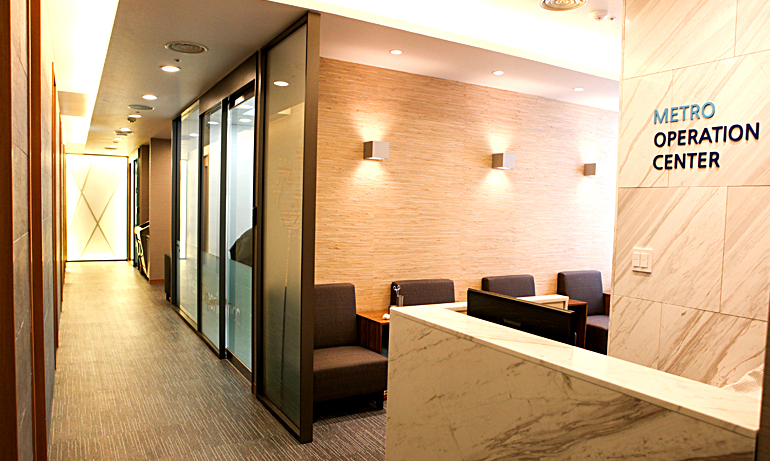
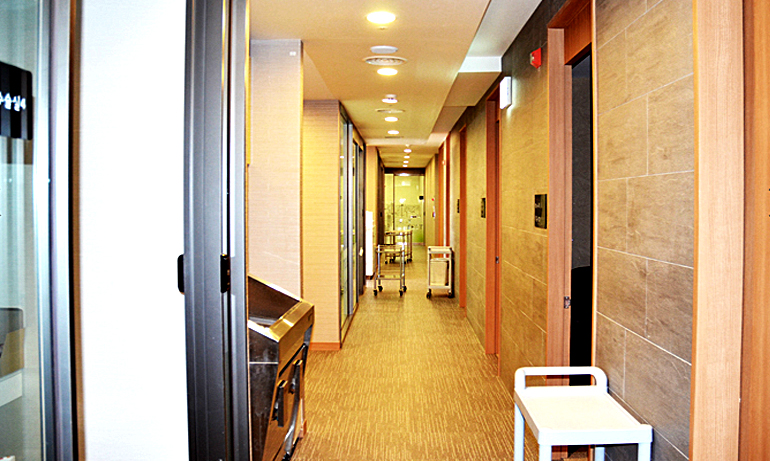
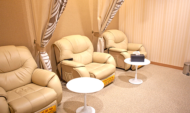
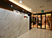
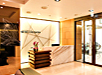
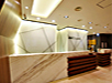
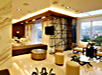
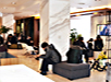
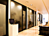
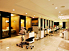
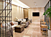
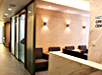
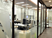
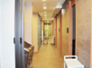
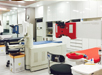
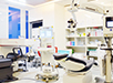
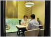
Have a quick look around Metro Eye Clinic


Surgery Reviews
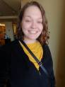
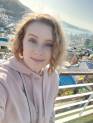
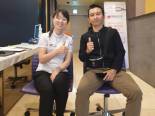
외국인 체험기

- 외국인 체험기

|
|
|
|
|
|
|
|
|
|
|
|

|
|
|
|
|
|
|
|
|
|
|